Secret Gardens
Glendale Community College student interns working behind the scenes are unearthing the hidden worlds of NHM’s Nature Gardens, plucking pollen from its greenery, tucking the fine powder under the Scanning Electron Microscope, and revealing the smallest bits of nature you’ve ever seen.

Published November 1, 2023
Jeremy Welch, a Glendale Community College student intern at NHM, is aiming for a career in environmental science. He is also a gardening enthusiast. So when Welch, along with another GCC intern, Katie Huguet Tuck, were invited to collect pollen from blossoming plants in the botanical showroom that is the museum's Nature Gardens, it was a welcome assignment. “I mostly just walked around the gardens looking for native things that either I liked or were pretty, or were just kind of cool plants…California poppies, our state flower, common Yarrow, goldenrod.”
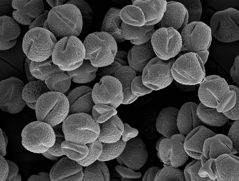
Collecting this essential ingredient of flora reproduction was part one of the assignment. Back in the lab, when they took the fine powder from each species of flower and delicately placed the material under the Museum’s Scanning Electron Microscope (SEM), another equally mesmerizing world emerged. SEMs have a more powerful magnification ability than traditional microscopes. Light microscopes use photons (particles of light) received by the human eye to let us see a magnified object, while SEMs employ electrons, negatively charged particles with short wavelengths interacting with the surface of that object, to create an image displayed on a computer screen; it can magnify much more than light microscopy can and expose very fine details.
Dr. Jann Vendetti, NHM’s Associate Curator of Malacology, who co-created the internship program, is excited to offer opportunities for students to do science in this indoor-outdoor museum.
“The beautiful plants of the Nature Gardens hold additional wonders in the form of pollen that are too tiny for us to see,” she said. “The SEM opens up this world, allowing us to appreciate their marvelous shapes." Looking at the gray SEM scans of the pollen while at the museum recently, interns Huguet Tuck and Welch found those geometries to be wondrous.
“My favorite is a poppy— it looks like an egg,” said Welch. “There are some awesome ones. I like the clarkia. It's got this weird triangular shape.” “I think that one looks like popcorn,” says Huguet Tuck, who is studying ecology and evolutionary biology.
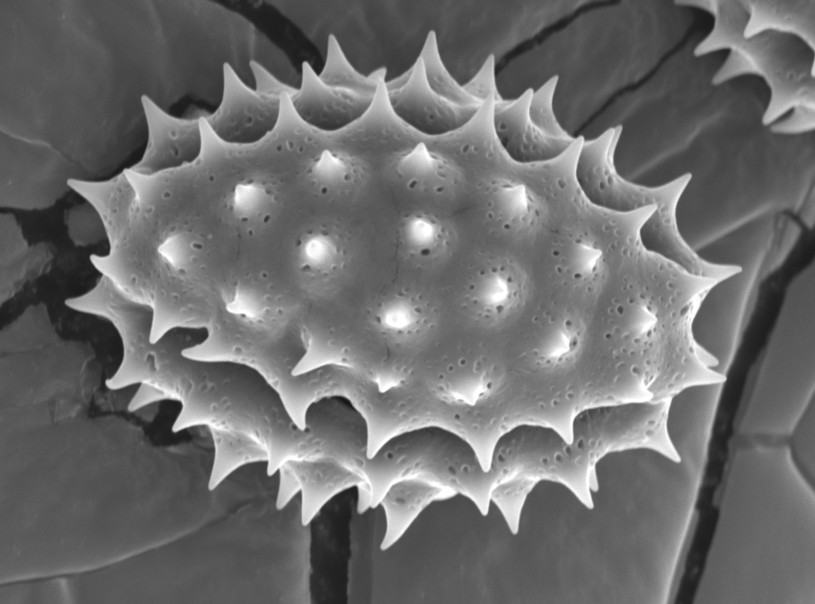
“That one [monkey flower pollen], looks like a volleyball. It's spiked.” Janice Lee, another intern, pipes in—“that looks like a melon.”
“That looks like a dragon fruit,” says Huguet Tuck of the elongated, prickly California goldenrod.

The fetching pink petals of the elegant clarkia, growing in the museum's Nature Gardens.


A monkey flower growing in the Nature Gardens

Robert Tarango, the fourth GCC intern working with Dr. Vendetti on Fridays this semester, is enthusiastic about the deep knowledge that can be gleaned from the 600 species of plants in the greenspace that visitors drink in daily.
“It is crazy how much diversity there is If you just go outside. Like me, every time I would come to the museum, it would be like, 'Oh, plants.' I wouldn't think much more beyond that,” Tarango said. “But, if you really actually look, you'll see how much of another world there is besides just the ones we happen to be passing by.”
With the SEM scans in hand, these four students are able to pose new questions about the posies: If they see similarities, could it be convergent evolution or are they closely related, which they can find out if they go even deeper to determine its DNA. The students say they may also be able to glean information from the scans about pollination techniques. In pollination, pollen grains are transferred—by wind, insects, birds, or other pollinators—from the male anther of a flower to the female stigma with the goal of producing seeds.
“We could look at the female parts to see if they attach easily, and look at advantages and disadvantages,” says Huguet Tuck. “It could be the dragon-fruit-looking pollen’s advantage is that it can attach really easily. Disadvantage? It can attach really easily to things that aren't important” to procreation.
Tiny Floaters, Big Ocean
Pollen aside, Tarango and Lee have been fixated on another SEM-assisted find: diatoms. They were working on a project looking at white abalone, Haliotis sorenseni, specimens donated to the Museum recently from a captive breeding program at UC Davis' Bodega Marine Laboratory (most abalone in California are highly endangered due to overfishing) and discovered that the mollusk had been fed diatoms. Diatoms are infinitesimally small algae made of silica that have lived in the world’s oceans for millions of years, and their glassy translucent bodies come in the most incredible shapes. These rectangles and triangles can be two microns long—about half as thick as spider silk!
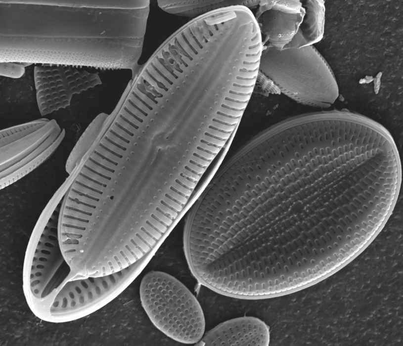
“It's cool because they literally looked like baskets just floating out and about,” said Tarango, who is studying marine biology. “They look very mechanical as opposed to biological.” Lee, who has a bachelor's degree in psychology, and worked in health care before going back to school to study biology, loves the fact that their college and the museum supports such a variety of natural history investigations.
“I think it really makes us have a broad view of everything. It’s a lot of fun doing all of these different things.” The students have unending praise for Vendetti, who also introduced the group to one of her favorite microscopic topics—the radula, the savage-looking mouthparts of snails.
“She's been nothing but supportive in helping us grow in our confidence in science and showing us how approachable everything is,” says Huguet Tuck. “Because it's so intimidating when you start something like this, especially if you've never done research before. But she is such a great mentor.”
“Dr. Vendetti is the type of person who will get excited over anything, and it's contagious,” said Lee. “She just has this zeal for everything in biology, and to be able to have a mentor who has that approach, that kind of attitude, I think is really powerful for us, too.”


