Think Small With SEM
Seeing the big picture with tiny specimens and one powerful scanning electron microscope

Try to imagine the smallest animal you’ve ever seen with the naked eye. Got it? Okay, what does its mouth look like?
Small morphological details like mouth shape can reveal exactly how the smallest creatures around are staying alive, and lets us get a better view of little things that can help us see the big picture.
Head Hunting Flies
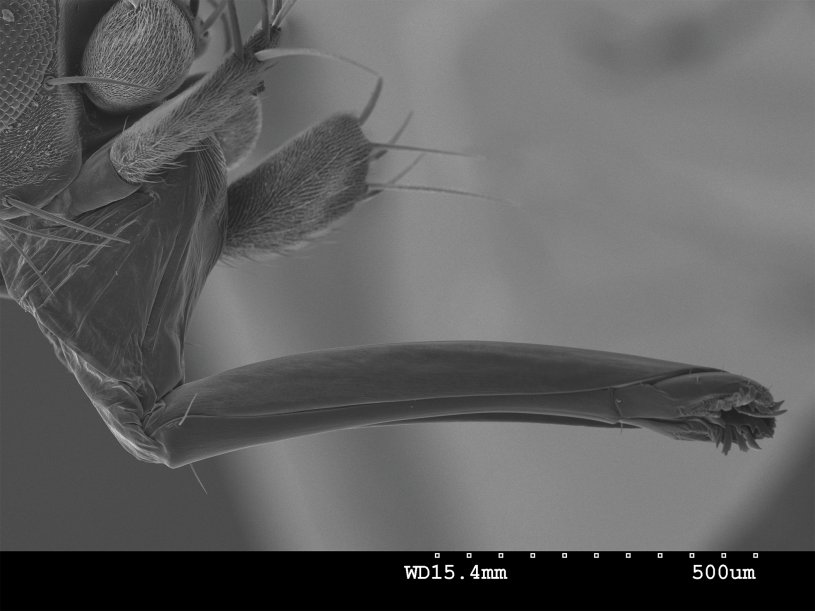
When NHM’s Entomology Curator Brian Brown and Collections Manager Giar-Ann Kung were doing fieldwork in Brazil, they noticed a phorid fly in the Dohrniphora longirostrata-group was leaving some ants headless. Kung suggested the ants were being decapitated, but Brown was initially skeptical. A closer look at the fly’s proboscis under NHM’s Scanning Electron Microscope revealed the structures that helped support Kung’s observation. Getting a closer look at the proboscis exposed nature’s tiny saw blade.
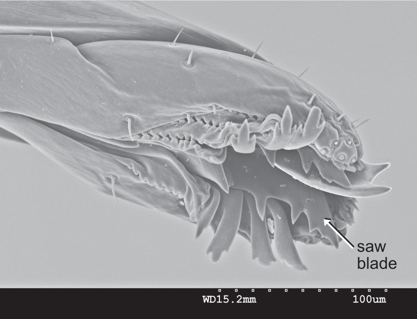
This is a SEM of a Dohrniphora longirostrata-group collected by Dr. Brian Brown and Giar-Ann Kung in Brazil, and a close up on its proboscis. This fly uses its proboscis with that serrated appendage to cut off the heads of its prey.
“When we can see more of the super small things, we get a better picture of the specimen; we get a new story to share.”
—Dr. Trina Roberts, Associate Vice President of Collections
A NEW LENS ON RESEARCH
The Scanning Electron Microscope has a more powerful magnification ability than other microscopes thanks to their use of electrons. While traditional microscopes use photons (particles of light) the SEM uses electrons, negatively charged particles that have much shorter wavelengths than photons. These shorter wavelengths help reveal much finer details. It’s like the first time you watched a sporting event televised in HD–suddenly you can see the blades of grass on the field.
This type of imaging has the power to help researchers understand their subjects on a deeper level, and the images they produce can help inspire wonder in our natural world. So when NHMLAC received a grant from the National Science Foundation to help fund the purchase of a new SEM, NHMLAC’s Research and Collections team was thrilled.
“The new SEM is an indispensable modern research instrument that will allow our researchers to view specimens and artifacts at tremendous magnifications and with superb resolution,” explains Dr. Jody Martin, Associate Vice President for Research. “Equally important, the SEM will be displayed for students and visitors to NHM to allow them to see 'science in action' and learn about the wonders of the microscopic world as they watch our scientists at work.”
HEADS UP
Ants are famously small, but under the magnifying power of the SEM, their stunning anatomy becomes clear. The head shapes, eye placement, and mouth structures of the ants below differ greatly from one another. Seeing such a high level of detail can point researchers in new directions of study. They're also just really cool.

Image by Giar-Ann Kung, Entomology Department
Even though it's big for an ant, getting this detailed a view of Notostigma carazzil would be impossible without a SEM.

Strumigenys elongata shows off its incredible head structure under the SEM.

Image by Giar-Ann Kung, Entomology Department
Atta cephalotes distinct heart-shaped head accommodates the large muscles needed to power the mandibles of this leaf-cutter ant

Image by Giar-Ann Kung, Entomology Department
Gigantops destructor's remarkably large eyes are on full display.
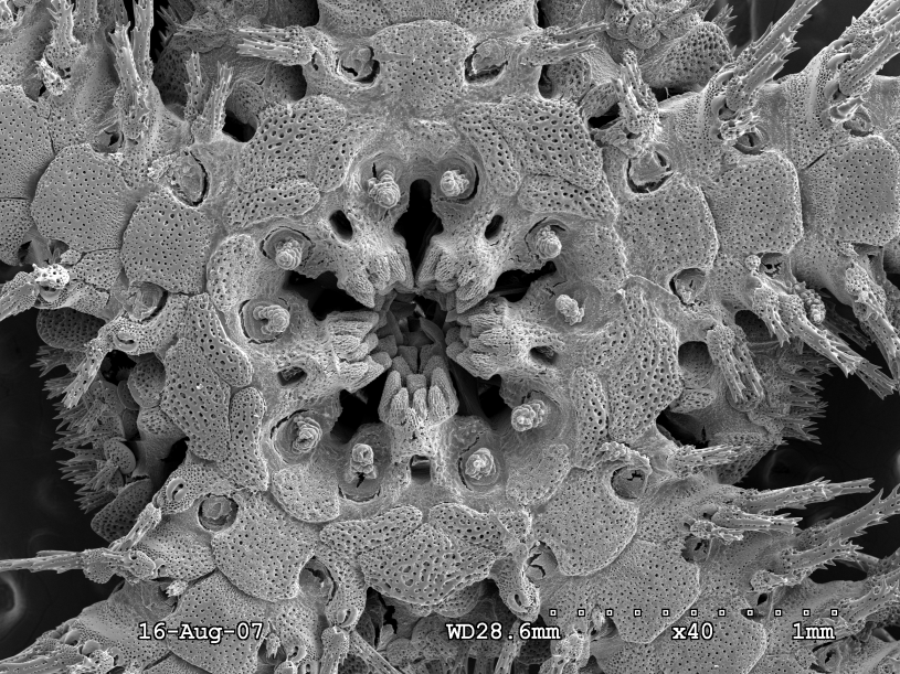
Whether it's the how of ant decapitation or helping scientists differentiate between minuscule mouths, SEM technology plays an indispensable role in Museum science while producing amazing images of the hidden, microscopic world all around us. The new SEM is up and running, and Museum researchers have been putting everything from minerals to marine worms under the microscope to see our natural world more closely than ever before. Be sure to check out NHM researchers getting small at the Scanning Electron Microscope Lab on your next visit.
