The museum will be closed on Thursday, November 28 for the Thanksgiving holiday. We will reopen on Friday, November 29.

Get the Scoop on the Scope

Zoom in on tiny specimens with the Scanning Electron Microscope.
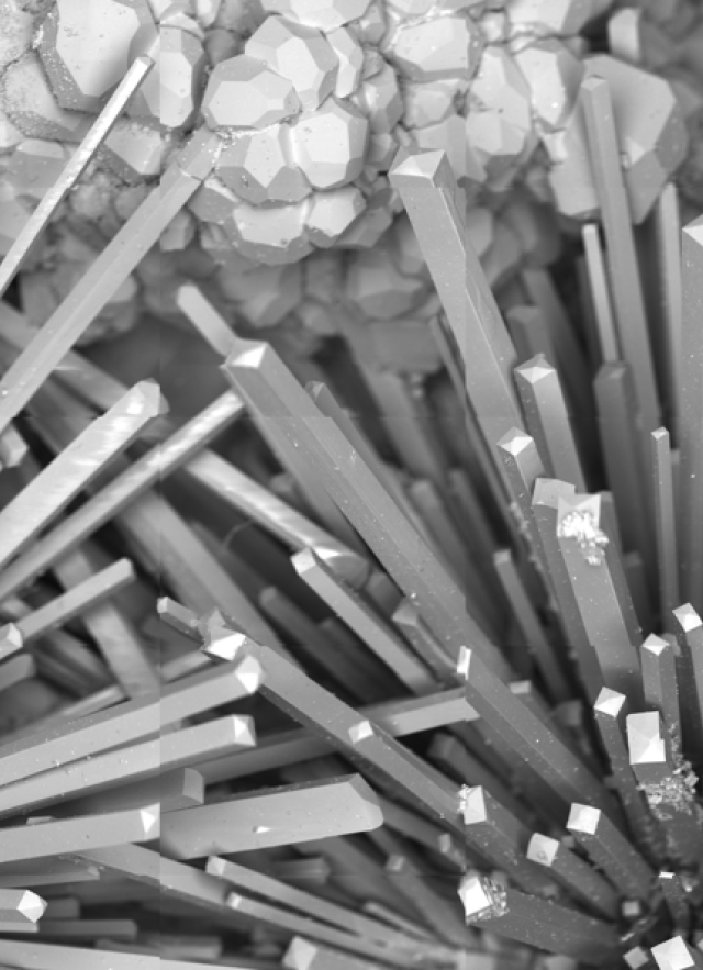
A black and white image of spiky mineral structures.

Lesson Plan by DeAnna Lee-Rivers, MA Ed.

suggested for 6 - 12th grade

Overview

Museum researchers use the Scanning Electron Microscope (SEM) to zoom in on tiny specimens and learn about worlds they otherwise wouldn’t be able to see. Now you too can view these worlds in the SEM Lab! Follow along with the provided worksheet to discover how the technology works, learn how a specimen is prepared for viewing, and meet some of the scientists using the SEM to advance their fields of research.

concepts

  • Scanning Electron Microscopes are powerful tools that work by directing a concentrated stream of electrons over the surface of a sample to create an image.
  • Magnification allows us to see an enlarged image of a specimen and increases the resolution: our ability to see details of a specimen or sample.
  • The SEM helps us to see objects at magnifications as low as 40x to as high as 100,000x! Light microscopes have a maximum magnification of only around 1,000x.
  • The Natural History Museum of LA County is unique in that it is an active research site–a place where scientists work to expand their understanding of the world.

objectives

  • Students will be able to explain how the SEM works.
  • Students will understand the scale of SEM magnification.
  • Students will be able to imagine what types of applications SEM technology has for the future.

duration

Pre-Visit: 30-50 minutes 

Visit: 20 minutes

Post-Visit: optional

location

Scanning Electron Microscope (SEM) Lab in the Gem & Mineral Hall

supplies

vocabulary

  • SEM
  • Electron
  • Resolution

Outline

  1. Facilitate a discussion with your class on the SEM and its applications by presenting the Pre-Visit Slides.
  2. During a trip to the Museum, explore the SEM lab and complete the worksheets.

  3. You may expand your learning with activities and conversation starters from the Post-Visit Slides and Post-Visit content at the end of this lesson plan.

Pre-Visit

A Scanning Electron Microscope (SEM) is a powerful instrument that uses electrons to create a detailed image of incredibly small specimens. Present the Pre-Visit Slides to your class to provide students with a foundation for their trip to NHM and exploration of the SEM Lab.

Visit

During a field trip to NHM, take some time to investigate the SEM Lab. You may split your class into small groups or have students work individually to complete the Get the Scoop on the Scope Worksheets. Note that there are two versions–A and B–which will allow your class to occupy the space efficiently.

Post-Visit

SEM technology has changed the way we look at the world and helped solved problems that we weren’t able to before. The best part is that we are able to imagine new possibilities for the future! Go through the Post-Visit Slides and consider the ideas below in your discussion on this question: How could SEM technology be useful for your community?

Here are some ideas:

  • Particulate matter and air quality - What size are the particles floating in our air? These particles (dust, soot, fibers, etc.) are considered pollutants and can get trapped in our lung tissue.
  • Disease diagnosis at cellular level - How could we use the SEM to investigate and diagnose diseases such as cancer?
  • Distinguishing between the good and bad bacteria in your body - Some bacteria can invade our cells and make us sick. How does microscope technology help us tell them apart?

Variation & Extension Ideas

In order to continue engaging with the exhibit and exchanging ideas, you will find additional resources below. Feel free to pick and choose which of these resources you would like to use.

Draw a picture of an organism or item that you would like to see under an SEM. Perhaps consider organisms living in or on our own bodies such as gut bacteria and face mites on skin.

Create-A-Creature: Choose an SEM image of an organism that looks especially interesting to you. Create a life story for that organism. Get as creative as you possibly can and use these questions to get started: 

  • What type of organism is it?
  • What does it eat?
  • Where does it live and how does it interact with other organisms?

Opportunities for Research: Further investigate a topic related to SEM that you find interesting. Prepare your findings in an essay or report. Here are some example topics:

  • Why do samples have to be dipped in gold? Why not another metal? 
  • Why do the cells have to be killed? 
  • Which scientific advancement or discovery made with the SEM is most interesting to you? Why?

Diversity in the field of SEM research: There are scientists from many different backgrounds who have contributed to SEM advancements and who use the technology currently. You can read about two of them below.
Research another scientist using microscopes in their field of study and present their biography to the class in the form of a poster.

  • Helio da Silva, Ph.D. (Universidade Federal Rural do Rio de Janeiro, Brazil) works on frog morphology, or  structure, and their place on the tree of life.  Part of his research involves investigating the structural characters overlooked over the years. He and his students use SEM to unravel details of tadpole "tooth" shape and development, sexual characteristics on the hands of frogs and toads, and warts on the skin of toads.
  • Itzahi Silva Morales, Ph.D. candidate (Departamento de Sistemática y Ecología Acuática, El Colegio de la Frontera Sur, Chetumal, Quintana Roo, Mexico) is one of the few biologists who conducts research on a group of marine animals known as peanut worms or sipunculans. She studies them with the use of SEM and has discovered, details in their morphology that have often been overlooked. Her research will greatly enhance knowledge of the diversity and evolutionary relationships of this group of worms.

Community Connections: How can YOUR community benefit from this technology? 

  • What would your community consider to be a benefit?
  • Might one community benefit from something different than another?
  • Based on what you learned from the SEM Lab and classroom discussions, how will you get your community members/leaders excited about this technology?

SEM Lab FAQs

Will my students be able to use the Scanning Electron Microscope during their visit?

  • No, the SEM is a powerful research tool used only by Museum staff who have specialized training and expertise. The SEM is always visible to Museum visitors through the window in the SEM Lab.

Will a scientist be demonstrating how the Scanning Electron Microscope works during our visit?

  • There may be a Museum scientist working with the SEM during your visit, but there is no scheduled programming. You will be able to look at the SEM from behind glass and learn how the SEM works through exhibit pictures and text.

Standards

NGSS

HS-PS4-5

S+E Practices

1, 5, 6, 8

Crosscutting Concepts

Influence of Science, Engineering, and Technology on Society and the Natural World (MS-ETS1-1, HS-ESS3-3)

CCSS ELA

WHST.9-12.2